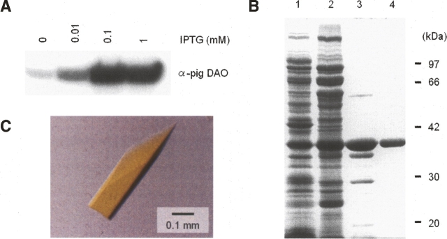
Figure 1.
Purification and crystallization of recombinant human DAO. (A) Western blot of recombinant human DAO produced in E. coli. (B) SDS-PAGE of purified DAO isolated from E. coli. The gel (10%) was stained with Coomassie blue. (Lane 1) Whole cell (containing 20 μg of protein), (lane 2) after heat treatment (59°C, 3 min) and 70% ammonium sulfate fractionation (20 μg of protein), (lane 3) DEAE Sepharose CL-6B column eluate (5 μg of protein), (lane 4) hydroxylapatite column eluate (2 μg of protein). Values indicate the molecular weight of the marker proteins: phosphorylase b (97 kDa), BSA (66 kDa), aldolase (42 kDa), carbonic anhydrase (30 kDa), and soybean trypsin inhibitor (20 kDa). (C) Crystal of human DAO.

