
Figure 2.
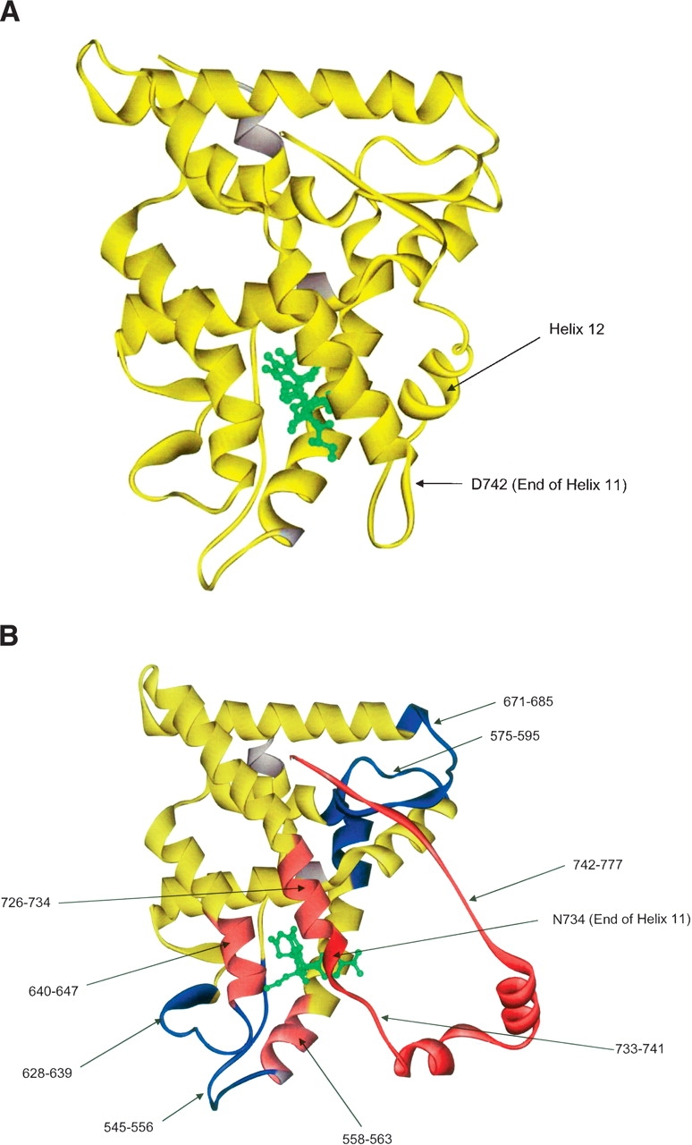
(A) The crystal structure of GR LBD with dexamethasone bound. This serves as a control for Figures 2B and 3A. (B) The crystal structure of GR LBD with RU-486 bound. Regions colored in blue show decreased exchange (increased protection) relative to GR LBD with dexamethasone bound. Regions colored in light red show a moderate increase in exchange (decreased protection), while regions colored in dark red show a strong increase in exchange (decreased protection). This experiment also serves as a control for Figure 3B.