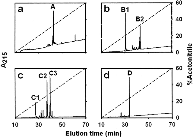
Figure 3.
RP-HPLC analysis of tryptic peptides present in each fraction separated after size-exclusion chromatography. Pooled fractions corresponding to each peak separated after chromatography on a Superdex Peptide HR 10/30 column (A, B, C, or D; see Fig. 2c) were sampled onto a C18 column (a–d, respectively). Elution was monitored by UV absorption at 215 nm (solid line). Major peaks belonging to each pooled fraction were numbered sequentially. The aqueous acetonitrile gradient is shown as a dashed line (see Materials and Methods).

