
Figure 1.
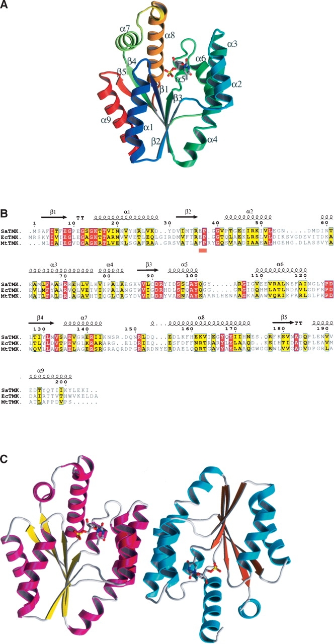
Structure of SaTMK. (A) Ribbon diagram of SaTMK with secondary structure marked and showing bound TMP. SaTMK is shown in rainbow colors from blue at the N terminus to red at the C terminus. TMP is shown in standard atom colors, with the carbon atoms in cyan. (B) Sequence alignment of SaTMK, EcTMK, and MtTMK. The secondary structure elements shown above the alignment are of SaTMK. The orange bar below the alignment indicates the conserved (E/F)P loop. (C) Ribbon diagram of the SaTMK dimer. The helices of the two molecules are shown in magenta and cyan, while the sheets are shown in yellow and orange. TMP is shown in standard atom colors, with the carbon atoms in gray.