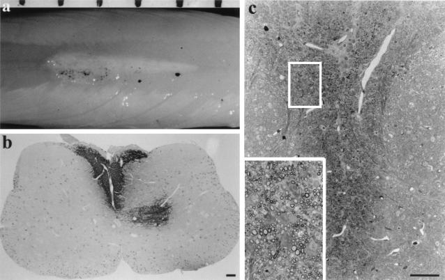
Figure 3.
Transplantation of oligosphere cells from a 16-month-old rat into md rats. Twelve to fourteen days later, a white streak of myelin was seen along the dorsal surface of the cord (a). The black dots are sterile charcoal marking the injection site. The space bar on top represents 1 mm. (b) Immunostaining of the transplanted cord showed PLP+ myelin in the dorsal funiculus with some myelin also appeared in the gray matter. Other areas of the spinal cord showed no PLP+ myelin except the PLP+ cell bodies. (c) Semithin sections stained with toluidine blue demonstrated that the dorsal funiculus was occupied by a large number of myelin sheaths. Inset is the enlargement of the boxed area in c. (Bar = 100 μm.)

