
Figure 8.
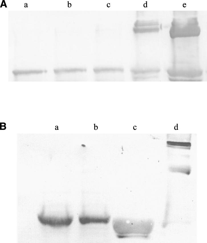
Western analysis of the Rel WT protein expressed in E. coli BL21 (DE3). (A) Rel WT protein probed with anti-Rel antibodies. Lanes a–c represent the crude cell lysate overexpressing Rel protein and treated as mentioned in Materials and Methods. Lanes d and e are the protein (control) run without DTT. (B) Rel WT protein probed with anti-His tag antibodies. Lanes a and b represent the crude cell lysate treated as mentioned in Materials and Methods. Lanes c and d represent the protein run in the presence and the absence of DTT, respectively, and act as controls.