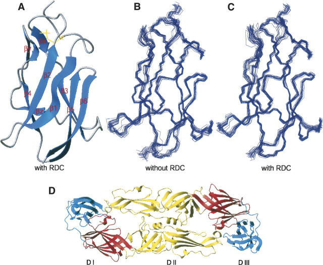
Figure 1.
Calculated structure of LGT-E-D3. (A) Ribbon display of the lowest-energy NMR structure of LGT-E-D3 calculated with RDC restraints. (B) Superimposition of 20 lowest-energy backbone structures calculated without RDC and (C) with RDC restraints, respectively. (D) Ribbon plot showing the three different domains of TBE envelope (dimer) protein: (red) domain I, (yellow) domain II, and (blue) domain III.

