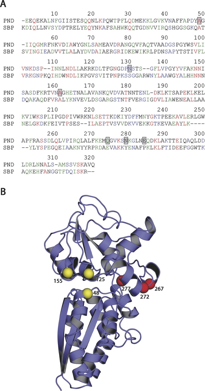
Figure 3.

(A) Sequence alignment of PhnD and E. coli sulfate-binding protein (SBP), positions of engineered cysteines are indicated by gray boxes. (B) Location of cysteine mutations mapped onto the crystal structure of SBP (PDB code 1SBP) using the sequence alignment to establish the correct register. Spheres indicate peristeric (yellow) and allosteric (red) positions where cysteine mutations were introduced. Note that PhnD contains several insertions relative to SBP; these amino acids are located in surface loops and are not included in the model shown here.
