
Figure 1.
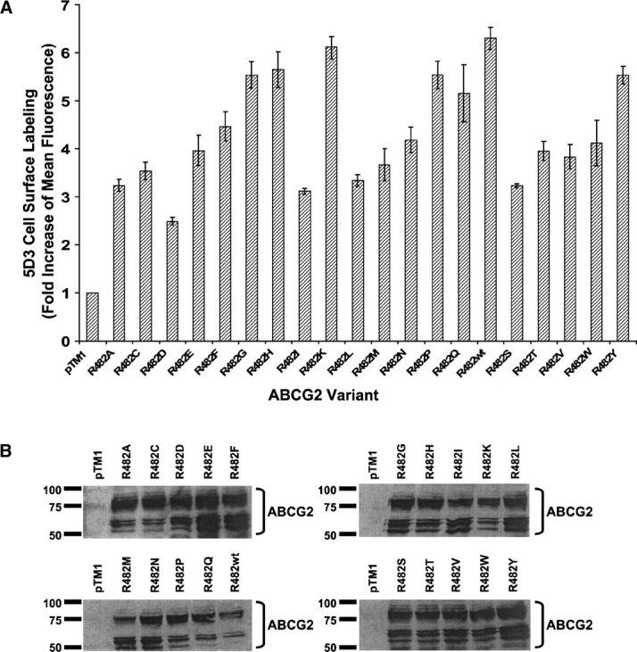
Expression and cell surface localization of R482X ABCG2 variants expressed in HeLa cells. (A) Cells were stained with the monoclonal antibody 5D3 (2 μg/200,000 cells) that recognizes a cell surface epitope of ABCG2. As the negative control, cells were transfected with the empty vector pTM1. The protein–antibody complex was visualized with a FITC-labeled secondary antibody (2.5 μg/200,000 cells) and the cells were analyzed for fluorescence by flow cytometry. Each bar represents the fold increase compared with the negative control cells transfected with pTM1, and X denotes any of the 20 standard amino acids. (B) Immunoblot analysis of cell lysates (20 μg protein), using the monoclonal antibody BXP-21 as described in Materials and Methods.