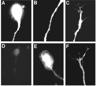
Figure 2.
TrkC immunoreactivity is detected at different neuronal segments. (A–C) Representative examples of neurons stained with antibodies to TrkC (Santa Cruz Biotechnology). The immunofluorescence signal is evident at the soma (A), along the axon (A and B); and at the growth cone (C). (D) Control experiment demonstrating specificity of staining. Preincubation of primary antibodies with the blocking peptide largely abolished the immunofluorescence signal. (E–F) Neurons were stained with antibodies to TrkC (Upstate Biotechnology); this antibody was raised against the extracellular domain of TrkC receptor. The immunofluorescence signal can be detected at the cell body and along the axon (E) and at the distal axon (F).

