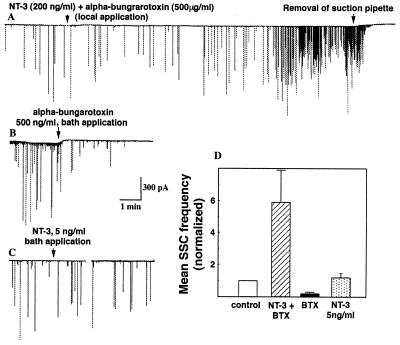
Figure 4.
“Leakage” of NT-3 from the superfused region does not contribute to the potentiation of ACh secretion at the preformed synapses. (A) Membrane currents recorded from the myocyte in the preformed synapse. NT-3 (200 ng/ml) together with α-bungarotoxin (500 μg/ml) was locally applied to the middle axonal segment as in Fig. 3A. The start of local perfusion is marked by the arrow. The postsynaptic myocyte was ≈400 μm away from the site of drug application. Withdrawal of the pipette used for the removal of the superfused solution (arrow) resulted in the accumulation of α-bungarotoxin in the dish medium and the inhibition of SSC at the preformed synapse. (B) Membrane currents recorded from an innervated myocyte in the preformed synapse. Bath application of α-bungarotoxin (500 ng/ml, marked by arrow) resulted in the decrease in the frequency of SSCs. (C) Membrane currents recorded from the innervated myocyte before bath application of NT-3 application (5 ng/ml, marked by arrow) and for a period of 15–18 min after NT-3 application (right trace). (D) Quantitative analysis of the data. In each experiment, the mean SSC frequency for a period of 15–18 min after the drug application was normalized to the mean SSC frequency before drug application. Each data point represents the mean ± SEM of seven to nine experiments.

