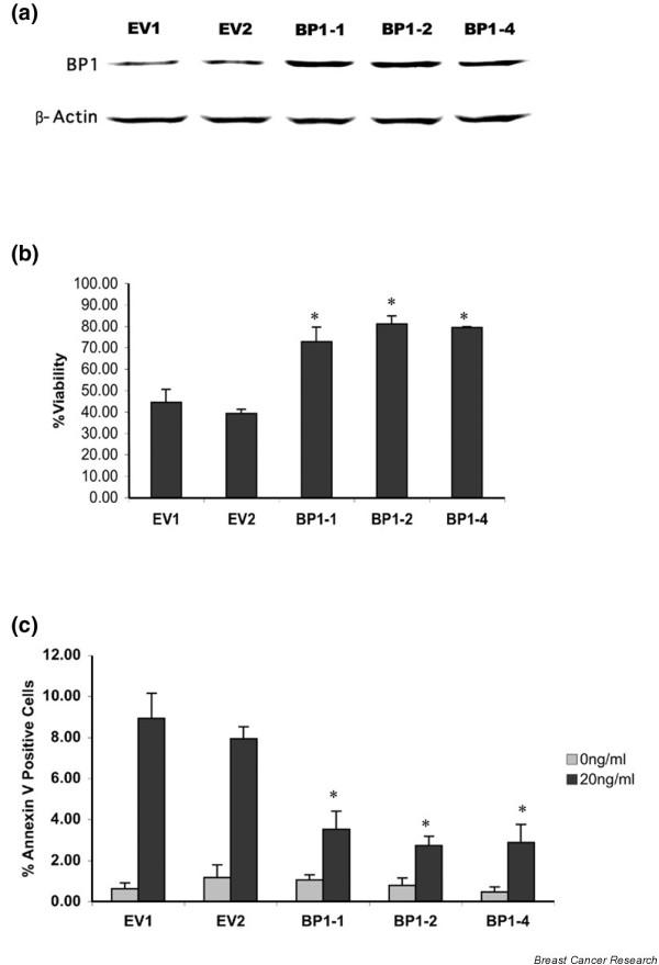
Figure 1.

Effect of BP1 on TNFα-induced cell death. (a) Western blot analysis of Beta Protein 1 (BP1) protein expression in MCF7/EV cell lines and MCF7/BP1 cell lines. (b) MCF7/EV and MCF7/BP1 cell lines were treated with 20 ng/ml TNFα for 72 hours. MTT assays were performed to assess cell viability. To calculate the percentage viability, absorbance values were compared in treated cells versus untreated cells. *Statistically significant differences (P < 0.0001). (c) MCF7/EV and MCF7/BP1 cell lines were treated with 20 ng/ml TNFα for 18 hours. Cells were labeled with both an Annexin V–FITC conjugate and propidium iodide to distinguish early apoptotic cells. Five fields of cells were photographed and counted for each sample in three independent experiments. The percentage of cells in each field with Annexin V staining in the plasma membrane, but showing exclusion of propidium iodide, is reported. *P < 0.0001.
