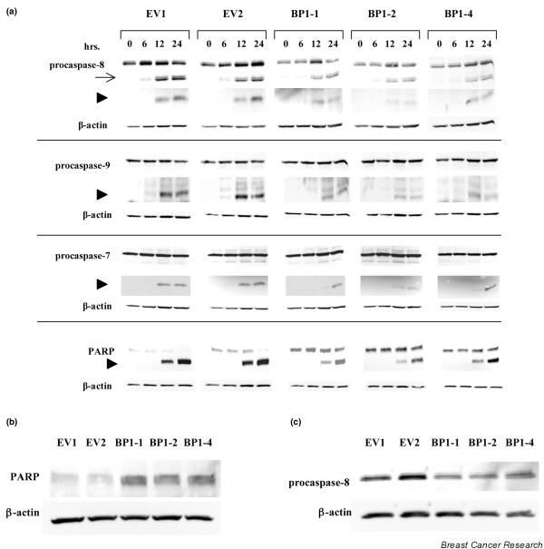
Figure 2.
BP1 inhibits TNFα-mediated caspase activation. (a) MCF7/EV and MCF7/Beta Protein 1 (BP1) cells were treated with 20 ng/ml TNFα. At the indicated times, protein was analyzed by western blot to examine the expression levels and processing of caspase-8, caspase-9, and caspase-7, as well as the substrate Poly(ADP-Ribose) Polymerase (PARP). In each case, the top band represents the uncleaved, inactive procaspase or full-length active PARP. Arrow, intermediate fragments; arrowheads, position of the expected cleaved product. (b) and (c) Western blot analyses of PARP and procaspase-8 expression in MCF7/EV and MCF7/BP1 cell lines.

