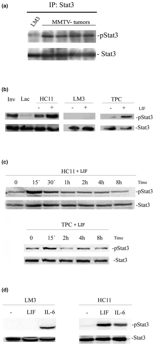
Figure 3.

Western blot analysis of phospho-Stat3 (pStat3) and Stat3. (a) Tumors growing in vivo: LM3 (lane 1) and different HIT transplants (lanes 2 to 5). (b-d) Cells growing in culture. (b) HC11, LM3 and tumor primary culture (TPC) cells treated with 80 ng/ml leukemia inhibitory factor (LIF) for 15 minutes; mammary glands at 48 hours of involution (Inv) and at fifth day of lactation (Lac) were used as positive and negative controls, respectively. (c) Time course of tyrosine phosphorylation of Stat3 (signal transduction and activators of transcription 3) in HC11 cells (upper panel) and TPC cells (lower panel) treated with 80 ng/ml LIF. (d) Tyrosine phosphorylation of Stat3 in HC11 and LM3 cells treated with LIF and IL-6 (both at 80 ng/ml) for 15 minutes.
