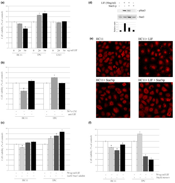
Figure 5.
Effect of LIF and CM on cell viability after 72 hours of treatment. Viability was assessed by crystal violet assays. (a) HC11, tumor primary culture (TPC) and LM3 cells were treated with leukemia inhibitory factor (LIF; 20 and 50 ng/ml). (b) HC11 and TPC cells were treated with 50% conditioned medium (CM) preincubated with or without LIF-neutralizing antibody. (c) HC11 and TPC cells were treated with 50 ng/ml LIF in the presence or absence of a Stat3 (signal transduction and activators of transcription 3) inhibitor peptide. (d, e) Effect of Stat3-specific inhibitory peptide (Stat3ip) on Stat3 phosphorylation levels (d) and nuclear translocation (e) induced by LIF. 400× (f) HC11 and TPC cells were treated with 50 ng/ml LIF in the presence or absence of PD98059. Data are percentages of internal control for each cell type (time 0) and are expressed as means ± SEM for four replicates. Experiments were repeated at least three times with similar results. Asterisk denotes statistical difference (P < 0.05) in a two-tailed Student's t test.

