Fig. 4.
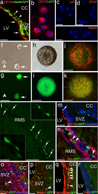
Contribution of transplanted CD133+ cells to the lineage of adult SVZ. (a) Confocal image of the SVZ region of adult Nestin/GFP mouse, stained with anti-CD133 (red). Arrows point to CD133+ ependymal cells. Arrowheads indicate Nestin-GFP cells in the SEZ. (Scale bar: 200 μm.) (b–e) Sorted CD133+ cells (red) from Nestin/GFP mice are not immunoreactive for GFP, LeX, GFAP, or Nestin during the first DIV. (Scale bars: b, 20 μm and c–e, 30 μm). (f–i) Clonally generated neurospheres from a CD133+ cell—isolated from Nestin/GFP mice—captured from the same area at 2 (f and g) or 6 (h and i) DIV with either bright field (f and h) or GFP (g and i) filters. Arrows point to Nestin/GFP+ spheres, whereas the arrowhead denotes a forming sphere with no GFP expression yet. (Scale bars: f and g, 75 μm and h and i, 100 μm.) (j and k) Differentiated spheres on coated surfaces at 5 DIV expressing Nestin/GFP and stained with LeX (red in j) or CD133 (red in k). (Scale bars: 100 μm.) (l) GFP+ cells (arrows) migrating along the RMS 2 weeks after transplantation of sorted CD133+/GFP− cells from Nestin/GFP mice into the lateral ventricles of wild-type-recipient mice. (Scale bar: 250 μm.) (Insets) Higher magnification of migrating GFP+ cells in the RMS. (Scale bar: 20 μm.) (m) A confocal photomicrograph of the SVZ region of a recipient mouse 1 week after the sorted CD133+ cells were injected into the lateral ventricle. Even in close proximity of the ventricular surface, GFP+ cells (arrow) are CD133− (red). (n–r) Migrating GFP+ cells (green) in the SVZ/RMS are colabeled with doublecortin (arrowheads in n), GFAP (arrowhead in o), Mash1 (arrowhead in p) or PSA-NCAM (arrowheads in q). None of the GFP+ cells are costained with Olig2 (r). CC, corpus callosum; LV, lateral ventricle; RMS, rostral migratory stream; SVZ, subventricular zone. (Scale bars: n–o, 250 μm.)

