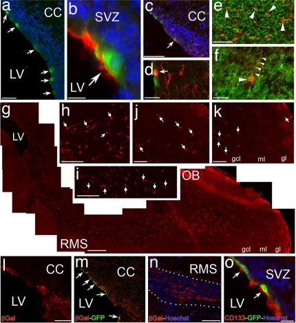
Fig. 5.
Generation of olfactory bulb interneurons by CD133+ ependymal neural stem cells in the postnatal forebrain. (a) GFP+ cells (green) that were transfected with an expression vector plasmid containing a short prominin-1 promoter (i.e., mP2) governing Cre-GFP expression line the ventricles (arrows). (Scale bar: 150 μm.) (b) A GFP+ transfected cell (arrow) in the ependymal layer is also CD133+ (red). (Scale bar: 15 μm.) (c and d) Fluorescent photomicrographs taken from Rosa26 reporter mice injected with mP2 plasmid. Transfected GFP+ cells (green) are in the ependyma whereas the β-gal+ cells (red) are in the ependyma and SEZ. Arrows point to transfected ependymal cells that started to express β-gal. (Scale bar: c, 200 μm and d, 50 μm.) (e) β-gal+ migrating cells (red) in the RMS are PSA-NCAM+ (green, arrowheads). (Scale bar: 75 μm.) (f) A migrating β-gal+ cell (red) in the layers of the olfactory bulb is TuJ1+ (green, arrowhead). Small arrowheads point to the process of the β-gal+ cell extending into the overlying layer of the olfactory bulb. (Scale bar: 50 μm.) (g) A photomontage of a P10 Rosa26 reporter mouse forebrain showing the entire extent of the RMS and olfactory bulb in a sagittal plane. The animal was injected with the mP2 construct at P3. β-gal+ cells (red) are distributed along the RMS from SVZ to the layers of the olfactory bulb. (Scale bar: 400 μm.) (h–k) Localization of β-gal+ cells in the RMS (arrows in h and i) and the layers of the olfactory bulb (arrows in j and k) of a P10 Rosa-26 reporter mouse. (Scale bars: 75 μm.) (l and m) A fluorescent photomicrograph of an adult Rosa26 reporter mouse forebrain injected with mP2 construct showing the SVZ region. β-gal+ cells (red) are distributed in the ependyma and SEZ, whereas transfected GFP+ cells line the ventricles (green, arrows). (Scale bar: 200 μm.) (n) Localization of β-gal+ cells (red) along the RMS en route to the olfactory bulb. Dotted white line outlines the borders of the RMS. (Scale bar: 250 μm.) (o) GFP+ transfected cells (green) in the ependymal layer of an adult Rosa26 reporter mouse are also CD133-immunoreactive (arrows, red). Hoechst staining (blue) was used to label nuclei. (Scale bar: 75 μm.) CC, corpus callosum; gcl, granule cell layer; gl, glomerular layer; LV, lateral ventricle; ml, mitral cell layer; OB, olfactory bulb; RMS, rostral migratory stream; SVZ, subventricular zone.

