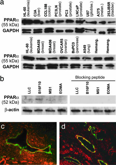
Fig. 1.
PPARα is expressed in tumor cells and endothelium of neoplastic tissues. (a) Western blot analysis of PPARα expression in cultured human tumor cells and hemangioma specimens. Nuclear extract from leukemia cells (HL-60) was used as a control. (b) Western blot analysis of PPARα expression in cultured mouse tumor cells. The specificity of protein expression was confirmed by abrogation by a PPARα-blocking peptide. GAPDH and β-actin levels were measured to demonstrate equal loading of protein in each lane. (c and d) Immunofluorescent double staining for CD31 and PPARα demonstrates PPARα expression in endothelium of human pancreatic cancer (BxPC3) in SCID mice (c) and in patient prostate cancer tissue specimens (d). CD31-stained endothelial cells are shown in green, PPARα-positive cells are red, and colocalization of the two colors are yellow. Colocalization of red and green fluorescence (yellow) indicates PPARα expression in blood vessels.

