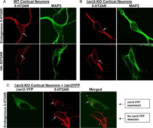
Fig. 2.
5-HT2AR localization in WT and β-arrestin-2-KO cortical neurons. (A) WT neurons. (B). β-arrestin-2-KO neurons. (Upper) Endogenous 5-HT2AR staining (Left, red) and MAP2 neuronal marker staining (Right, green). (Lower) Live cell HA-594 Alexa Fluor antibody staining of neurons transfected with an N-terminally tagged HA-5-HT2AR. Expression profiles were quantified by counting neurons based on robust, weak, or absent membrane staining. WT: 55 of 369 had weak staining, 2 of 369 had robust surface staining, and 312 of 369 had no discernable surface staining. KO: 57 of 411 had weak staining, 333 of 411 had robust surface staining, and 21 of 411 had no discernable surface staining. (C) β-arrestin-2-KO neurons were transfected with β-arrestin-2-YFP (βarr2-YFP) and stained for endogenous 5-HT2AR [shown as βarr2-YFP (Left, green), 5-HT2AR (Center, red), and merged image (Right)]. Note the localization of the endogenous 5-HT2AR on the cell surface of the nontransfected neuron, compared with the internalized receptors in the neuron expressing β-arrestin-2-YFP as indicated.

