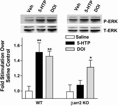
Fig. 5.
Agonist-induced ERK1/2 phosphorylation in frontal cortex of WT and β-arrestin-2-KO mice. Frontal cortex was dissected 15 min after vehicle (saline), 5-HTP (100 mg/kg, i.p.) or DOI (1 mg/kg, i.p.) DOI treatment, as described in Fig. 1. Brain lysates were resolved and analyzed by Western blot and densitometry as described in Fig. 4. The serotonin precursor (5-HTP) significantly stimulated ERK1/2 in the frontal cortex of WT, but not β-arrestin-2-KO mice; DOI stimulated ERK1/2 phosphorylation in both genotypes (saline vs. drug, **, P < 0.01; *, P < 0.05). One-way ANOVA performed within each genotype, followed by Bonferroni post hoc analysis. Data are the mean ± SEM (n = 9–13 mice per genotype per treatment). A representative blot of P-ERK and T-ERK is shown.

