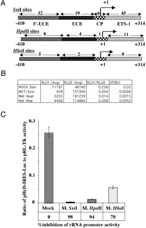
Fig. 3.

A, location of the recognition sites of different methylases on human rRNA promoter. B, human rRNA promoter activity in HepG2 cells transfected with pHrD-IRES-Luc, where the rRNA promoter region was methylated with M.SssI, M.HpaII, or M.HhaI and AdoMet. 1.5 × 10⁵ cells were transfected with 500 ng of mock-methylated or methylated plasmid DNA and promoter activity was analyzed as described in the legend to Fig. 2B. Table B represents the average value of the luciferase activities and the ratio of RLU1 (pHrD-IRES-Luc) to RLU2 (pRL-TK) from each set of transfected cells. C, graphical representation of the data.
