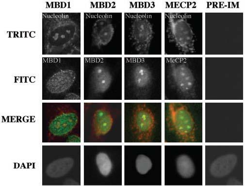
Fig. 5. Colocalization of methyl-binding proteins with nucleolin in HepG2 cells.

Co-localization studies were performed with antibodies specific for methyl-binding proteins raised in rabbit (except MBD2, which was raised in sheep) and monoclonal antibody (C23) against nucleolin, a nucleolus-specific protein. MBDs were recognized by fluorescein isothiocyanate-conjugated secondary antibody (green fluorescence), and nucleolin was recognized by TRITC-conjugated secondary antibody (red fluorescence). The yellow signal indicates co-localization of MBDs with nucleolin.
