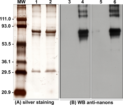
Figure 2. Silver-Stained SDS-PAGE and Western Blot Analysis of Nanon Proteins.
(A) Silver staining of 2.5 μg (lane 1) or 5 μg (lane 2) of nanon extract subjected to 10% SDS-PAGE.
(B) Western blot performed with two distinct mouse anti-nanon antibodies (1:2,500). Lanes 3 and 5, pre-immune sera; lane 4, mice 1; lane 6, mice 2. Molecular weight markers (MW) are on the left side.

