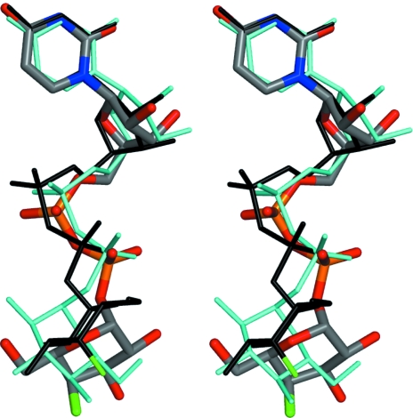
Figure 5.
Overlay of two UDP-FGal molecules observed in the complex with TbGalE (this work) and EcGalE (PDB code 1uda; Thoden et al., 1997 ▶) together with UDP-Glc from the complex with human GalE (PDB code 1ek6; Thoden et al., 2000 ▶). The UDP-FGal bound to TbGalE is depicted as in Fig. 2 ▶. The ligand from the bacterial enzyme complex is shown as black sticks with green to mark the F position; UDP-Glc is drawn as cyan sticks.

