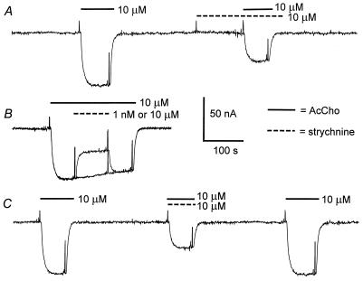
Figure 1.
Block of AcCho-current by strychnine. (A) Control current evoked by AcCho in an oocyte expressing neuronal α2β4 AcChoRs. After 3 min, the oocyte was superfused with strychnine alone, and then the AcCho-current was elicited again in the continuous presence of strychnine. (B) Superimposed records of AcCho-current showing the effects of 1 nM and 10 μM strychnine. (C) Control current, then simultaneous application of AcCho plus strychnine, and the recovered AcCho-current. The records were obtained from the same oocyte. For this and subsequent figures, the membrane was voltage-clamped at −60 mV, and the timings of drug applications are indicated by continuous bars for AcCho and dashed bars for strychnine above the records and by brief depolarizing pulses used to monitor membrane conductance changes. Inward currents are denoted by downward deflections of the trace.

