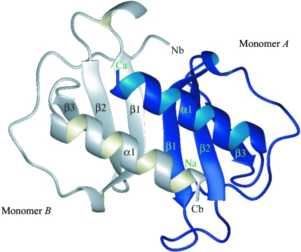
Figure 2.
Ribbon diagram of the overall three-dimensional structure of the dimer of human MIP-3α. The dimer is composed of a six-stranded β-sheet and two parallel α-helices. β-Strands are shown as arrows and α-helices as coils. This figure was generated with the program MOLMOL (Koradi et al., 1996 ▶).

