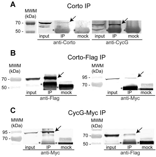
Figure 4. Corto and CycG interact in vivo.
A. CycG co-immunoprecipitates with Corto in embryonic extracts. Protein extracts from 0–14 h embryos were incubated with rabbit anti-Corto antibodies (IP) or rabbit preimmune serum (mock). Western blot analysis was performed using rat anti-Corto antibodies (left) or rat anti-CycG antibodies (right). Note specific precipitation of Corto (70 kDa) and the 68 kDa CycG species (arrows). The asterisks label unspecific IgG signals in all panels. B. Myc-CycG co-immunoprecipitates with Corto-Flag in S2 cell extracts. S2 cells were co-transfected with pAct::Corto-Flag and pAct::Myc-CycG. Proteins extracts were incubated with either mouse anti-Flag antibodies (IP) or mouse anti-HA antibodies (mock). Western blot analysis was performed using mouse anti-Flag antibodies (left) or mouse anti-Myc antibodies (right). Specific precipitation of Corto-Flag (left) or Myc-CycG (right) is indicated by arrows. C. Corto-Flag co-immunoprecipitates with Myc-CycG in S2 cell extracts. S2 cells were co-transfected with pAct::Corto-Flag and pAct::Myc-CycG. For immunoprecipitation we used either mouse anti-Myc antibodies (IP) or mouse anti-HA antibodies (mock), and for detection, mouse anti-Myc antibodies (left) or mouse anti-Flag antibodies (right). Specific precipitation of Myc-CycG (left) or Corto-Flag (right) is indicated by arrows. 4% of the starting material used in each IP (input) and 50% of the immunoprecipitated material were loaded onto the gel in all our assays.

