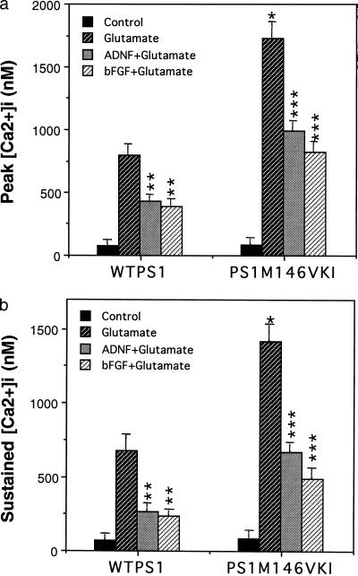
Figure 2.
Hippocampal neurons from PS1M146VKI mice exhibit enhanced calcium responses to glutamate: suppression by bFGF and ADNF. Hippocampal cultures from wild-type mice WTPS1 and PS1M146VKI mice were pretreated for 24 h with saline (control), 100 ng/ml bFGF, or 0.1 pM ADNF9. The [Ca2+]i was then monitored before and after exposure to 50 μM glutamate. Cumulative data for the peak [Ca2+]i (a) and the sustained [Ca2+]i (5 min after glutamate; b) following exposure to 50 μM glutamate. Values are the mean and SE of determinations made in 4–6 cultures (10–18 neurons analyzed per culture). ∗, P < 0.01 compared with the value for WTPS1 cells exposed to glutamate. ∗∗, P < 0.01 compared with the value for WTPS1 cells exposed to glutamate alone. ∗∗∗, P < 0.01 compared with the corresponding value for WTPS1 cells and to the value for PS1M146VKI cells exposed to glutamate alone. Probability values were determined by ANOVA with Scheffe’s post hoc tests.

