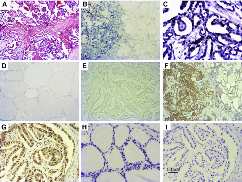
Figure 2.
In situ hybridization of parvovirus B19 DNA and IHC staining of B19 VP1/VP2 antigen in PTC. (A) Typical histopathological features of PTC. (B and C) The tumour cells of PTC showed strong nuclear staining by ISH, but the staining was almost negative in epithelia in tumour-adjacent tissue (B) and normal thyroid tissue (D). (E) Negative control (B19 probe was substituted by linearized pBR328 DNA). (F and G) Immunohistochemistry staining was for B19 VP1/VP2 antigen in PTC specimen and positive signals were seen in the cytoplasm of many malignant thyroid epithelia of PTC, but not in tumour-adjacent tissues. (H) No expression of VP1/VP2 antigen in normal thyroid samples. (I) Negative control (the primary antibody was replaced with mouse IgGI). Original magnification; × 40 (A, B and F); × 200 (C–E, G–I).

