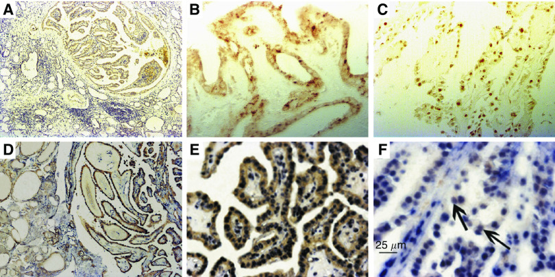
Figure 3.
Immunohistochemical staining of NF-κB p65 and double labelling of NF-κB p65 and B19 DNA in the samples of PTC. (A) NF-κB immunoreactivity in malignant tissues of PTC, but not in tumour-adjacent tissues. (B) Immunoreactivity of NF-κB in the cytoplasma of a PTC sample. (C) Nuclear translocation of NF-κB in a PTC sample. (D) NF-κB IHC and B19 DNA ISH double staining signal in malignant epithelium in PTC, but the staining intensity was much weak in epithelia in tumour-adjacent tissue. (E) Cytoplasmal expression of NF-κB p65 protein and robust nuclear presence of B19 DNA in identical tumour cells. (F) Coexpression of NF-κB p65 protein and B19 DNA in the nuclei of the tumour cells of PTC (black arrow). Original magnification: × 40 (A and D); × 200 (B, C and E); × 400 (F).

