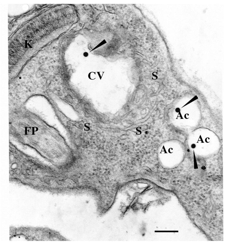
Figure 1. The contractile vacuole of Trypanosoma cruzi.

An epimastigote observed by transmission electron microscopy. Notations are flagellar pocket (FP), acidocalcisomes (Ac), kinetoplast (K), contractile vacuole bladder (CV), tubules forming the spongiome (S). Note that similar electron dense material (closed arrowheads) is observed in both the CV and the acidocalcisomes. Scale bar = 0.25 μm. Similar features were also observed in amastigote and trypomastigote forms. Reprinted with permission from Montalvetti et al., 2004.
