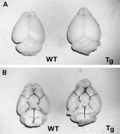
Figure 3.
Dorsal (A) and ventral (B) views of brains from WT and Tg mice. No gross anatomical difference was detected between the two sets of mice (n = 4 each). Intracardiac 5% India black injection revealed no anatomical abnormalities of the circle of Willis between the two groups (B, n = 4 each). Magnification: ×4.

