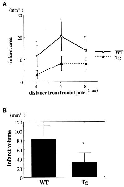
Figure 6.
Infarct volumes in Tg and WT mice. Infarct size was analyzed 24 hr after MCA occlusion using 2,3,5-triphenyltetrazolium chloride staining. (A) Infarct areas for three coronal sections from rostral to caudal are shown. Significant differences were found in Tg and WT mice (n = 9 each). (B) Infarct volume was smaller in Tg mice than in WT mice (n = 9 each). Data are expressed as means ± SD. Statistical analysis is performed by using Student’s t test; ∗, P < 0.01, ∗∗, P < 0.05)

