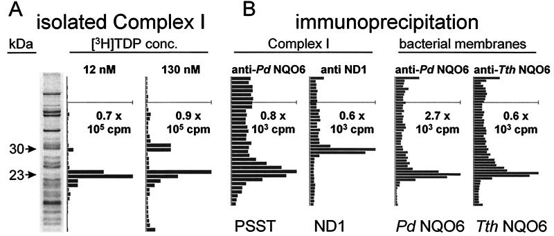
Figure 3.
Identification of the photoaffinity-labeled complex I high-affinity binding site protein (molecular mass 23 kDa) as PSST and the 30-kDa protein as ND1 and of the photoaffinity-labeled bacterial NDH-1 site protein (molecular mass 23 kDa) as NQO6. (A) SDS/PAGE analysis as in Fig. 2A but complex I isolated from ETP by blue native PAGE after labeling. Two of the darkest protein bands at 52 and 56 kDa are from complex V. (B) Immunoprecipitation of labeled complex I involved blue native PAGE followed by treatment with anti-Pd NQO6 antibody for PSST and anti-bovine ND1 antibody for ND1. Immunoprecipitation of labeled bacterial membranes involved direct treatment of P. denitrificans and T. thermophilus preparations with anti-Pd NQO6 antibody and anti-Tth NQO6 antibody, respectively.

