
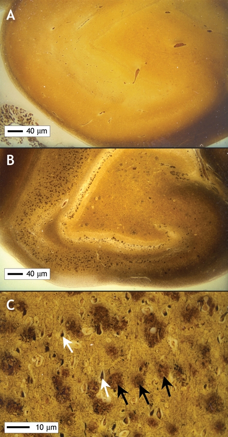
Figure 2: Images of normal hippocampus (A) and hippocampus of a patient with Alzheimer disease (B) [Bielschowsky stain]. The numerous dark brown spots seen in the abnormal hippocampus are the neuritic plaques typical of Alzheimer disease. At higher magnification (C), these plaques (black arrows) and tangles (white arrows) seen in Alzheimer disease are clearly visible. Image by: Images courtesy of Dr. Ian R.A. MacKenzie, Department of Pathology, University of British Columbia