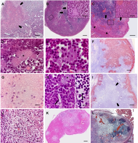
Figure 4. Illustrations of some of the criteria used for scoring.
Panels A–E, G, H and J–L display HE stained sections, panels F and I show sections immunostained with Y. pestis specific antibodies. In the text below, the criteria are referred to according to the numbering in Table 1. A: Wedge shaped abscess (criterion 4) indicated by arrows. Within the abscess bacterial colonies are visible as pink patches. B: Peripheral layer of PMNs containing bacterial foci (criterion 9), with a blunt demarcation (arrows) from the lymph node tissue (criterion 11). Inset: higher magnification to show the characteristic horseshoe shaped nuclei of the PMNs, and a bacterial focus. C: Layer of PMNs bordering a peripheral band of bacteria, cell debris and PMNs (criterion 10). The inset shows the typical PMN morphology of the cells within the layer. Behind this layer, bacterial aggregates are seen as pink areas (arrowheads) containing purple dots that are, as seen as higher magnification (not shown here) PMNs and cell debris. D: Patch of densely packed bacteria, bordered by PMNs (criterion14). Packed bacteria form a pink 8-shaped area at the centre of the picture (star). E: Atypical bacterial patch (criterion 15). At the center of the picture an aggregate of bacterial rods, not as densely packed as the preceding one, is loosely surrounded by inflammatory cells. F: A large bacterial zone (criterion 17), stained brownish on the preparation. G: Isolated host cells within a bacterial zone (criterion 19). Isolated host cells and cell remnants are seen amid a sea of bacteria which gives a “ground glass” appearance to this part of the LN section. H, left: bacterial infiltration around host cells (criterion 20). A bacterial strand (arrow), that seems to originate from a nearby colony (star), passes between host cells. H, right: Free floating bacterium, indicated by an arrow (criterion 21). I: Zone of reduced tissular density (arrows) outside bacterial areas, which are brownish on this preparation (criterion 26). This image also shows flame-like inward bacterial projections (criterion 18). J: Area of reduced host cell density with a reticular pattern (criterion 27) and containing numerous pycnotic cells (criterion 32). K: Moth eaten appearance (criterion 33) of a lymph node with areas of contrasting tissular densities. L: Vascular congestion (criterion 40), showing bright red on this preparation. Magnifications: Panels A–C, F, I–L : bar = 200 µm. Insets of panels B and C: bar = 50 µm. Panels D, E, G and H: bar = 10 µm.

