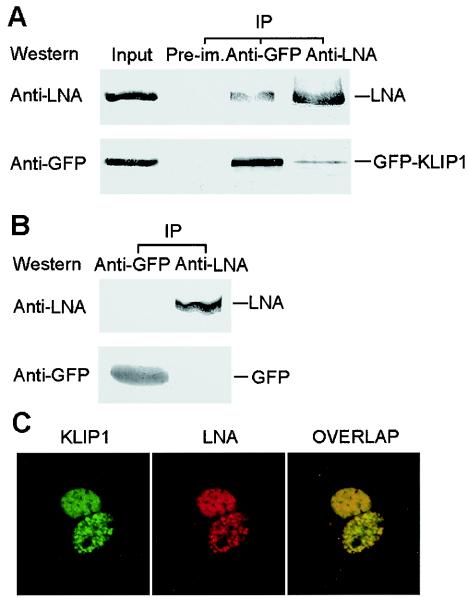
FIG. 8.
KLIP1 interacts with LNA in vivo. (A) Lysate from GFP-KLIP1-transfected BCBL-1 cells was immunoprecipitated with either anti-GFP (lane 3) or anti-LNA (lane 4) antibodies and then subjected to Western blotting with these two antibodies, respectively. The cell lysate was also precipitated with preimmune serum (lane 2, denoted Pre-im.) as a negative control. Lysates that were equal to 35% of those used in immunoprecipitation were run in PAGE and detected by Western blotting with anti-GFP and anti-LNA antibodies (lane 1). (B) Reciprocal coimmunoprecipitation was performed with GFP-transfected BCBL-1 cells to verify the specificity of the interaction between LNA and KLIP1. (C) Speckle green and red fluorescence distribution was observed in the nucleus of COS-7 cells transiently transfected with GFP-KLIP1 and pDsRed-LNA plasmid DNA. The distribution of both KLIP1 and LNA fusion proteins was identical and overlapped with each other, indicating colocalization of both proteins.

