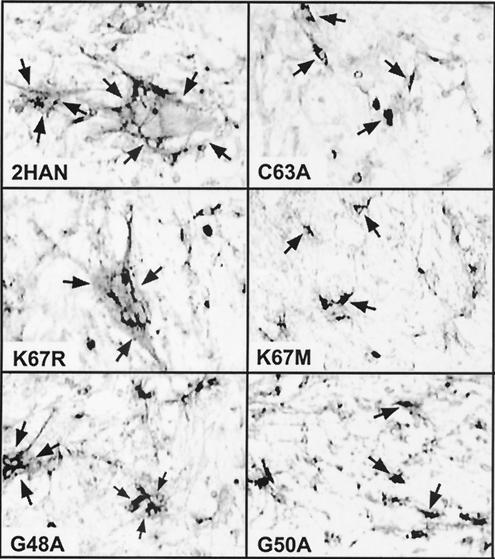
FIG. 2.
Syncytium-inducing activity of p10 constructs. Cells were transfected with various p10 constructs and fixed at 36 h posttransfection. The fixed cells were immunostained with anti-HA monoclonal antibodies and goat anti-mouse IgG conjugated with alkaline phosphatase. Arrows in the left panels indicate the boundaries of antigen-positive syncytial foci. The brighter areas surrounded by darkly stained areas indicate antigen-positive cytoplasm surrounding multiple antigen-negative nuclei in a single syncytium. Arrows in the right panels indicate single, darkly staining antigen-positive cells in monolayers transfected with syncytium-negative p10 constructs. Magnification, ×100. A subset of p10 constructs are shown, which include substitutions that did not significantly alter the fusogenic activity of p10 (K67R), those that drastically reduced both the number and size of syncytia (G48A), and those that eliminated p10-induced syncytium formation (C63A, K67 M, and G50A). The qualitative fusion activities of all of the p10 constructs, assessed in a similar manner, are summarized in Fig. 1C.

