FIG. 6.
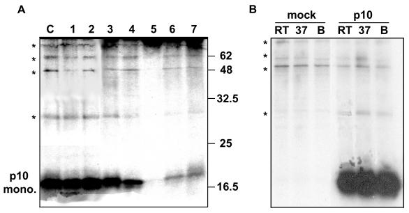
Cross-linking analysis of p10. (A) Radiolabeled cell suspensions were prepared from transfected cells expressing the p10-2HAN construct. The suspended cells were treated with one of seven different chemical cross-linking reagents (lane 1, PEO; lane 2, DTME; lane 3, DSS; lane 4, DST; lane 5, EGS; lane 6, DSP; lane 7, NHS). Control cell suspensions (lane C) were mock-treated with cross-linkers. The cells were disrupted, and the presence of p10 monomers or multimers was detected by SDS-PAGE under nonreducing conditions following immunoprecipitation with anti-HA antibody. The relative migration of molecular mass markers (in kilodaltons) is indicated on the right. Asterisks indicate the locations of background cell proteins present in all samples, including the control. (B) Mock- or p10-transfected cells were treated as described for the cross-linking control in panel A. Samples were treated at room temperature (RT), 37°C, or boiling (B) prior to gel analysis. *, locations of the faint high-molecular-weight protein species, indicative of background precipitation of host cell proteins rather than multimeric species of p10.

