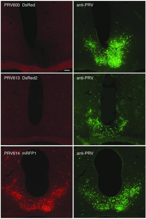
FIG. 2.
Coronal sections through the SCN, illustrating a red fluorescent signal at 96 h after inoculation of the right eye with strain PRV-Bartha recombinants. The left panels illustrate the expression of red fluorescent reporters in SCN-infected neurons. The right panels illustrate the same sections stained with an anti-PRV antibody and a secondary antibody conjugated to Alexa-488 (green). Note the absence of a red fluorescent signal in strains PRV600 (top left panel) and PRV613 (middle left panel), whereas virtually all PRV614-infected SCN cells produced a red fluorescent signal (bottom left panel).

