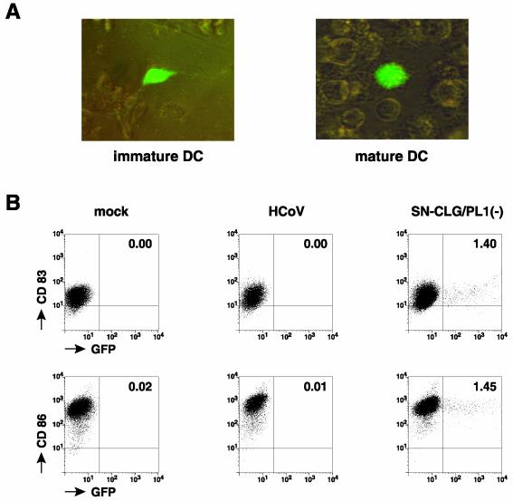
FIG. 5.
vec-CLG-mediated GFP expression in human DCs. (A) GFP expression was analyzed by fluorescence microscopy of immature and mature DCs transduced with supernatant SN-CLG/PL1(−). (B) Flow cytometry analysis of CD83, CD86, and GFP expression of mature DCs that have been either mock infected, infected with HCoV 229E, or transduced with supernatant SN-CLG/PL1(−). Indicated values represent the percentage of green fluorescent cells.

