
Figure 4.
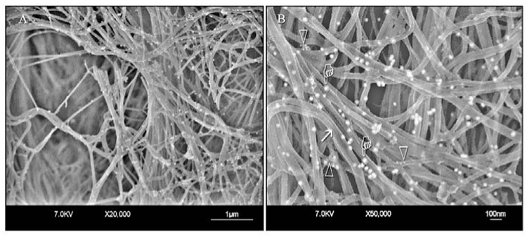
FEISEM micrographs of the collagen fibrillar network in normal hard dentin after immunolabeling. (A) Low magnification. (B) High magnification. Collagen fibrils appear unmodified, with surface cross-banding features (arrow) and gold nanoparticles (pointers) along the fibrils. Gold nanoparticles specific for proteoglycans appear as clusters of smaller electron-lucent particles around the collagen fibrils (open arrowheads). These clusters can be seen only at high magnification.