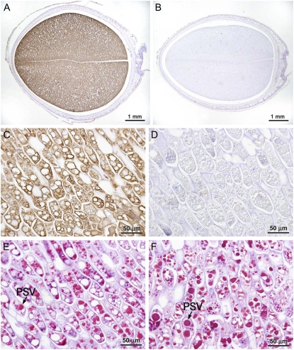
Figure 3.
Distribution of CAPPA antigen in immature (R6) CAPPA line cotyledons. A and B, Total cotyledon sections of CAPPA (A) and progenitor cultivar ‘Jack’ (B) immunostained with anti-APPA serum and counterstained with haematoxylin. C and D, Photomicrographs of a similarly treated longitudinal cotyledon section of CAPPA (C) at higher magnification and a control section treated with preimmune serum (D). E and F, Parallel sections of CAPPA and ‘Jack’ were stained with Schiff's reagent to observe the anatomical features.

