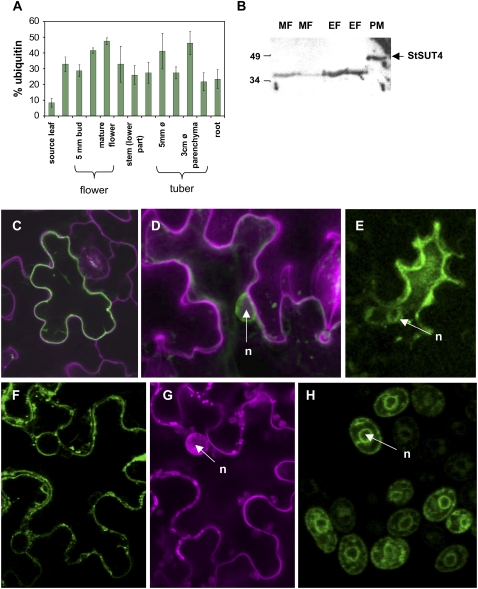
Figure 1.
A, Expression pattern of StSUT4 in sink and source organs as determined by real-time PCR. StSUT4 expression increases during flower development and strongest expression is detected in young developing tubers and mature flowers. B, Western-blot analysis of StSUT4 in leaves of potato. The microsomal fraction (MF) has been loaded in the first two lanes. Plasma membranes (PM) and endosomal membranes (EF) have been separated by two-phase partitioning and loaded on SDS-PAGE. In each lane, 15 μg of membrane proteins are loaded. StSUT4-specific peptide antibodies (Weise et al., 2000) detected the StSUT4 protein in the correct size of 47 kD only in the plasma membrane fraction. C, D, F, and G, Expression of StSUT4-GFP fusion expressed under the cauliflower mosaic virus 35S promoter in a pCF203 derivative in A. tumefaciens-infiltrated tobacco leaves. E, The same StSUT4-GFP construct expressed in infiltrated potato leaves. C, F, and G, Single scans. D and E, Overlay projections of confocal z stacks. GFP is not only detectable at the plasma membrane, but also in a perinuclear ring as shown by propidium iodide staining. F, StSUT4-GFP fluorescence is detectable at the plasma membrane of tobacco cells as well as in perinuclear rings. G, Same cell shown in F with propidium iodide-specific filter settings. H, Yeast cells expressing a LeSUT4-GFP construct under control of the Adh1 promoter in the yeast expression vector 112A1NE. GFP fluorescence is detected at the plasma membrane and in ER stacks surrounding the nucleus. n, Nucleus.

