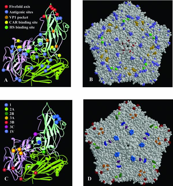
FIG. 3.
(A) Ribbon diagram showing the secondary structural elements of a viral protomer of SVDV (VP1, light blue; VP2, light green; VP3, pink). The location of the amino acid changes between CVB5 and SVDV (SPA/2/'93) is shown as differently colored spheres and explicitly indicated. (B) Space-filling model of an SVDV pentamer subunit showing the disposition of substitutions on the external part of the capsid colored as in A. (C) Ribbon diagram of an SVDV protomer highlighting the amino acids contributing to the seven antigenic sites residues as different colored spheres and explicitly indicated. (D) Space-filling model of an SVDV pentamer subunit showing the disposition of the antigenic sites colored as in C.

