Fig. 2.
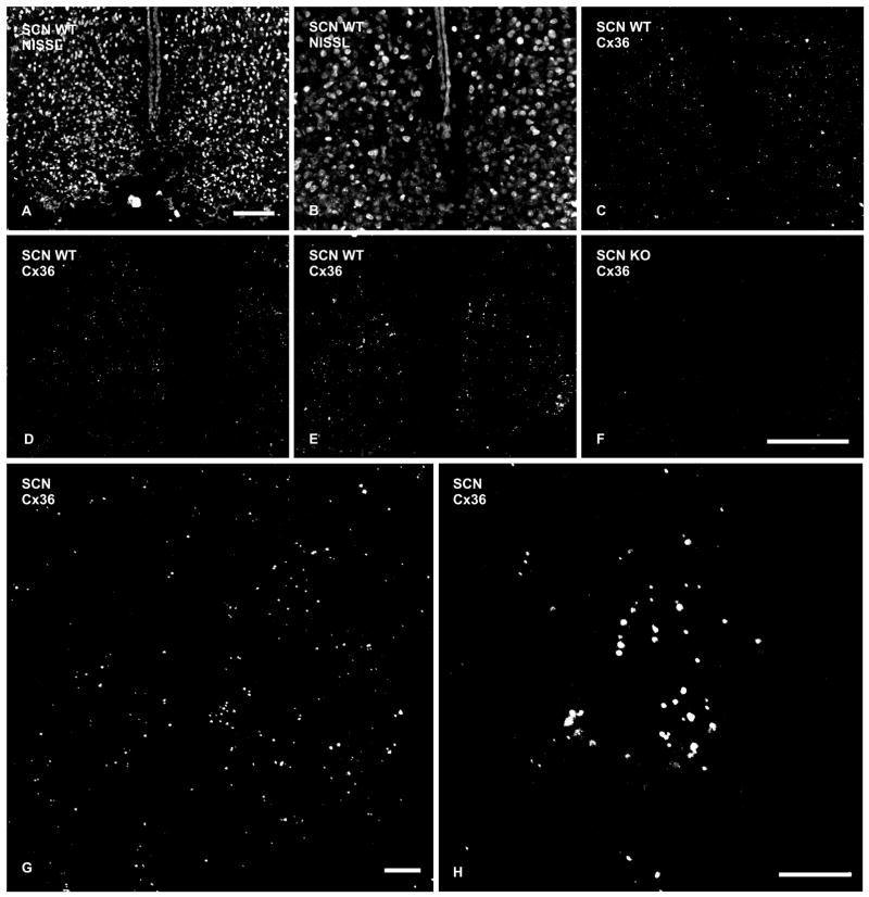
Immunofluorescence labeling of Cx36 in the SCN of adult mouse. (A) Low magnification bilateral view of Nissl-stained SCN. (B,C) Higher magnifications of the same field within a caudal level of the SCN Nissl-stained with red fluorochrome (B), and showing fine, punctate labeling for Cx36 (C). (D,E) Similar fields of the SCN as in B, showing punctate immunolabeling for Cx36 at midlevel through the nucleus (D) and at a rostral level (E). (F): Similar field of the SCN as in B and C, except from a Cx36 knockout mouse showing an absence of immunofluorescence puncta. (G,H) Confocal immunofluorescence showing Cx36-positive puncta in a z-stack of 10 sections through 6 μm of the SCN, and higher magnification of a cluster of puncta in the dorso-medial region of the SCN. Scale bars: A, 100 μm; B–F, 100 μm (shown in F); G,H, 10 μm.

