
Fig. 3.
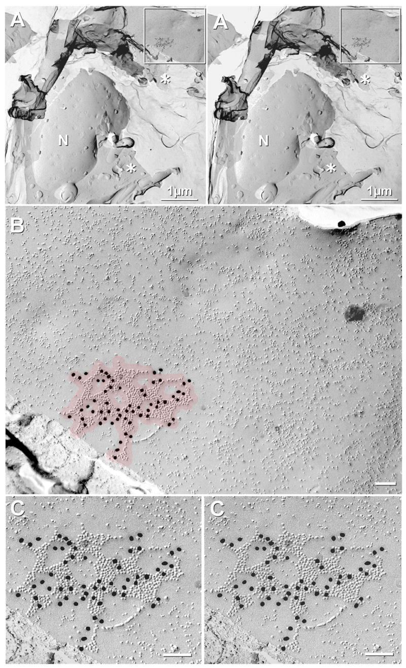
Low magnification to high magnification stereoscopic views of Cx36-labeled gap junction on plasma membrane of neuronal cell body in ventromedial aspect of SCN in a sample that was doubled-labeled for Cx36 (18-nm gold beads) and Cx32 (6 nm and 12 nm gold beads, none present in this image; but see Fig. 6C–E). (A) Stereoscopic image reveals the continuity of the cytoplasm (asterisk), as maintained by the secondary carbon support film applied before removal of the Lexan support layer (inset shown at higher magnification as B,C). The neuronal nucleus (N) has ca. 45 nuclear pores. (B) Overview of immunogold labeled “reticular” gap junction in broad expanse of neuronal plasma membrane. Red overlay indicates the 28-nm “zone of probable labeling” for connexons (Kamasawa et al., 2006). Note the absence of non-specific “background” labeling. Connexons are 9–10 nm, whereas other IMPs range from 4–8 nm in diameter. (C) High-magnification stereoscopic image of Cx36-labeled gap junction. Sixty gold beads label 622 connexons [Labeling efficiency, LE = 1:10 (Rash and Yasumura, 1999)]. N = nucleus; * = cytoplasm.
In this and subsequent FRIL electron micrographs, calibration bars are 0.1 μm, unless otherwise indicated.