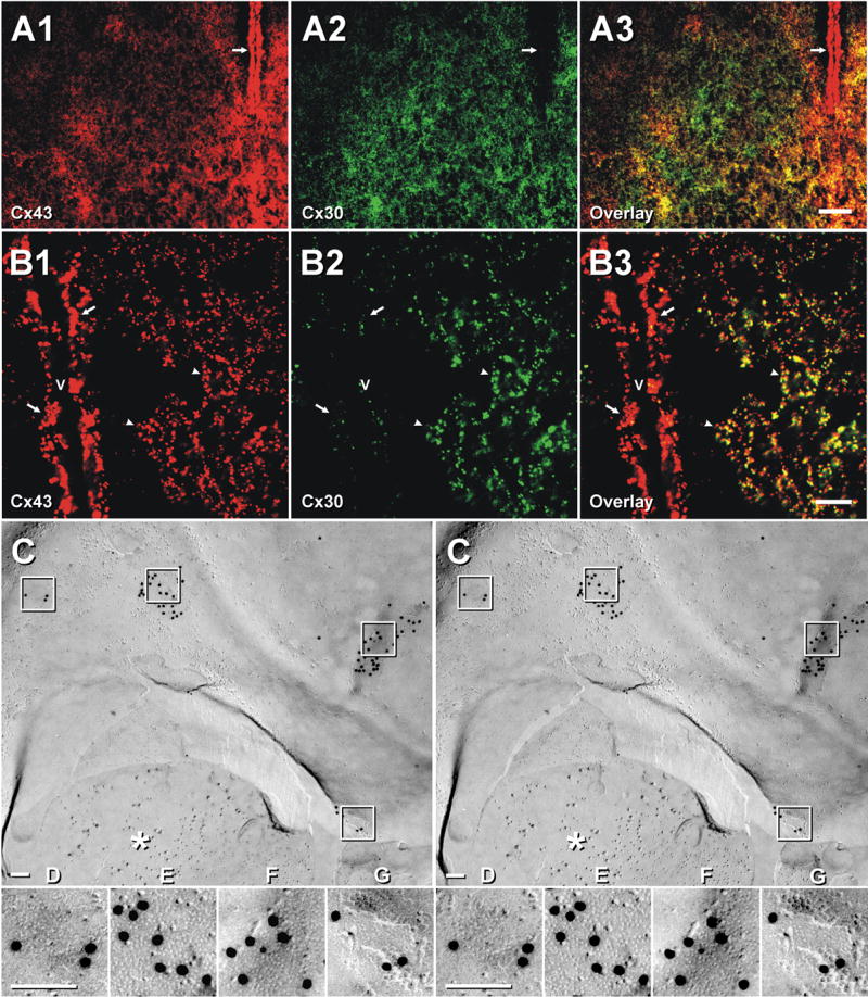
Fig. 7.

Immunofluorescence and FRIL labeling for Cx43 and Cx30 in the SCN and adjacent hypothalamic areas of adult mouse (A,B) and rat (C–G). Double labeling for Cx43 (A1) and Cx30 (A2) in a adult mouse SCN. Higher densities of Cx43- and Cx30-immunopositive puncta are seen within the SCN than in adjacent hypothalamic areas (A1–A3). Ependymal cells lining the third ventricle contain abundant Cx43 (A1 arrow) but lack Cx30 (A2, arrow). (B) Confocal immunofluorescence of the SCN showing co-localization of Cx43 (B1, arrowheads) and Cx30 (B2, arrowheads) as seen in overlay (B3, arrowheads). Ependymal cells are intensely labeled for Cx43 (B1, arrows) but are sparsely labeled for Cx30 adjacent to neuropil (B2, arrows). v, third ventricle. Scale bars: A, 50 μm; B, 10 μm. (C–G) Oligodendrocyte-to-astrocyte gap junctions in FRIL replica of SCN after immunogold labeling for Cx43 (20-nm gold beads) and AQP4 (10-nm gold beads). asterisk = GFAP filaments in astrocyte cytoplasm. Oligodendrocyte plasma membrane E-face has four gap junctions, portions of which are inscribed and shown in higher magnification stereoscopic images (D–G), which reveals the characteristic close spacing of E-face connexon pits. One gap junction (G) reveals the fracture step from oligodendrocyte E-face to astrocyte P-face. All E-face labeling of gap junctions is to cytoplasmic determinants of connexons in the underlying astrocyte plasma membrane (Massa and Mugnaini, 1982; Mugnaini, 1986; Rash et al., 1997, 2001b).
