
Fig. 8.
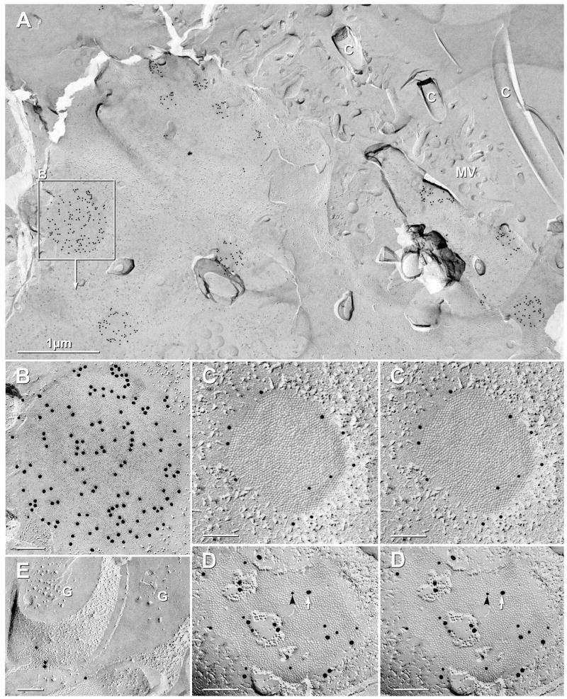
Ependymocyte and astrocyte gap junctions labeled for Cx43 vs. Cx30. (A,B) In a small area of a FRIL sample double-labeled for Cx43 (20-nm gold) and AQP4 (10-nm gold beads), 16 gap junctions linking lateral margins of ependymocytes were well labeled for Cx43. C = cilia; MV = microvilli. Inscribed area shown at higher magnification as (B). (B) Ependymocyte gap junction E-face heavily labeled for Cx43. (C–D) In stereoscopic images of ependymocyte-to-ependymocyte gap junctions double-labeled for Cx43 and Cx30, gap junctions were labeled only for Cx43 (C; 10-nm gold) but not for Cx30 (20-nm gold; none present), whereas gap junctions between astrocytes (D) were labeled for both Cx43 (10-nm gold, black arrowhead) and Cx30 (20-nm gold, white arrow). (E) Sub-ependymal astrocyte gap junction labeled for Cx43. From the same sample as Fig. 7C and 8A–B. G = GFAP filaments in astrocyte cytoplasm.