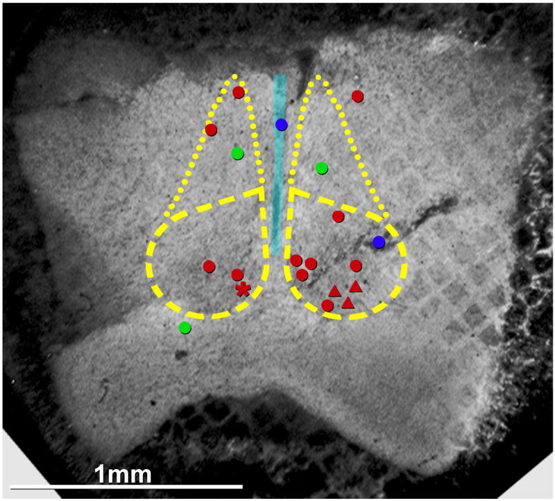
Fig. 9.
Composite confocal grid map of Cx36-labeled gap junctions on photomapped SCNs, including one on neuronal cell body (red asterisk) and 11 on dendrites (red dots) of adult, plus three on dendrites of P14 (red triangles). Left side of image illustrates locations of gap junctions in a single replica that was double-labeled for Cx36 plus Cx32 (red asterisk, Fig. 3; and red dots, from Figs. 4A–B designate neuronal gap junctions; green dots designate oligodendrocyte gap junctions, from Fig. 6C–E). Right side of image presents data compiled from five replicas from adult and one replica from P14. Red triangles = neuronal gap junctions from P14, Fig. 5; blue dots = astrocyte gap junctions (lower blue dot, Fig. 8E) and ependymocyte gap junctions (upper blue dot, Fig. 8A,B); dashed lines = SCN; dotted lines = subparaventricular extension of the SCN (Saper et al., 2005).

